


Ultrasound, also known as sonography, is an imaging method that uses high-frequency sound waves to produce real-time images of the body. Ultrasound is increasingly being used by Sports Medicine physicians, Rheumatologists, Orthopedists, and Primary Care doctors in performing evaluations and injections of different muscles, tendons, ligaments, nerves, and joints. With the advancement of this technology, ultrasound machines have become smaller and more portable. This has allowed treating clinicians to be able to use real time, point of care ultrasound, to assist in the diagnosis and treatment of their patients. It is also used for many different types of injections.
The use of ultrasound improves the accuracy of the injection of corticosteroids, hyaluronic acid, and other therapies such as Platelet Rich Plasma, Prolotherapy or Stem Cells. Ultrasound can also be used for joint. Lastly, guided injections can be used diagnostically to help determine which structures are generating the patient’s pain.
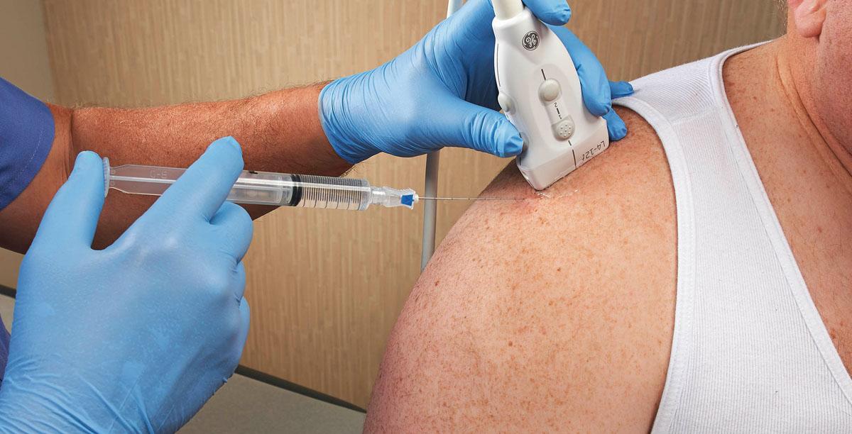
Ultrasound-guided injections allow the practitioner to visualize the needle in real time as it enters the body. This assures that the medication is accurately injected at the intended site. Despite good intentions, even in the most experienced hands, blind (injections performed without imaging) injections are not 100% accurate and, in some joints, accuracy is as low as 30%-40%. With ultrasound guidance the accuracy of nearly every joint injection exceeds 90% and, in many cases, approaches 100%. Additionally, ultrasound guided injections have been shown to be less painful than injections based on palpation alone.
Although there are many different types of imaging that can be used to assist with injections, ultrasound has a few distinct advantages.
1) Ultrasound has no radiation.
2) Ultrasound allows us to visualize the bony joint as well as the surrounding structures.
3) Ultrasound can identify fluid better than conventional radiographs and can see fluid that may have accumulated in and around joints, tendons, muscles, nerves and other soft-tissue structures.
Knee: Ultrasound can help evaluate a variety of structures within the knee including the quadriceps and patellar tendons, the extra-articular ligaments (MCL, LCL, etc.), and some meniscus injuries. It can also be used to see if there is fluid within the knee joint.
Hip: Hip joint injections may be performed for osteoarthritis of the hip and the diagnosis and management of labral tears. Imaging is nearly always used when performing injections into the hip joint due to the deep location of the joint and the proximity of blood vessels and nerves. It is estimated that blind injections are accurate 50% to 80% of the time. Ultrasound allows us to visualize the hip joint, bursa, muscles and tendons surrounding the hip. The use of ultrasound when performing a hip injection increases the accuracy to up to 96%.
Shoulder: Studies have shown that ultrasound of the shoulder is just as sensitive and specific as MRI in the diagnosis of rotator cuff injury. Ultrasound can facilitate the more accurate injection of multiple structures in the shoulder including the Acromioclavicular (AC) joint, the Glenohumeral joint, the biceps tendon, and the subacromial bursa.
Recent Comments